Content Status
Type
Linked Node
MGIT 960 Instrument: Principle for Culture
Learning Objectives- Understand the fundamental principles underlying the MGIT 960 instrument for tuberculosis (TB) culture.
- Learn about the system's automated culture method, which relies on fluorescence detection.
H5Content
Content
Mycobacteria Growth Indicator Tube (MGIT) contains:
- 7.0 ml of modified Middlebrook 7H9 broth base
- Growth supplement essential for the growth of Mycobacterium tuberculosis complex
- MGIT PANTA, antibiotic mix to prevent contamination
- Oxygen-quenched fluorochrome (tris 4, 7-diphenyl-1, 10-phenonthroline ruthenium chloride pentahydrate) embedded in silicone at bottom of the tube
Principle
- During bacterial growth within the tube, free oxygen is utilized and replaced with carbon dioxide.
- With the depletion of free oxygen, the fluorochrome is no longer inhibited, resulting in fluorescence within the MGIT tube.
- The intensity of fluorescence is directly proportional to the extent of oxygen depletion.
- Inoculated MGIT tubes are either incubated at 37ºC, read manually under ultra violet light or incubated into MGIT 960 instrument and monitored for fluorescence
- Positive and negative results (Figure 1 and 2)
- Growth of mycobacteria increases fluorescence; at the time of positivity, there are approximately 105-106 Colony-forming Units (CFU) per ml of medium
- The instrument declares a tube negative if it remains negative for six weeks (42 days)
- The detection of growth in the medium is also visually observed for the:
- Presence of non-homogeneous light turbidity, or
- Small granular/ flaky appearance
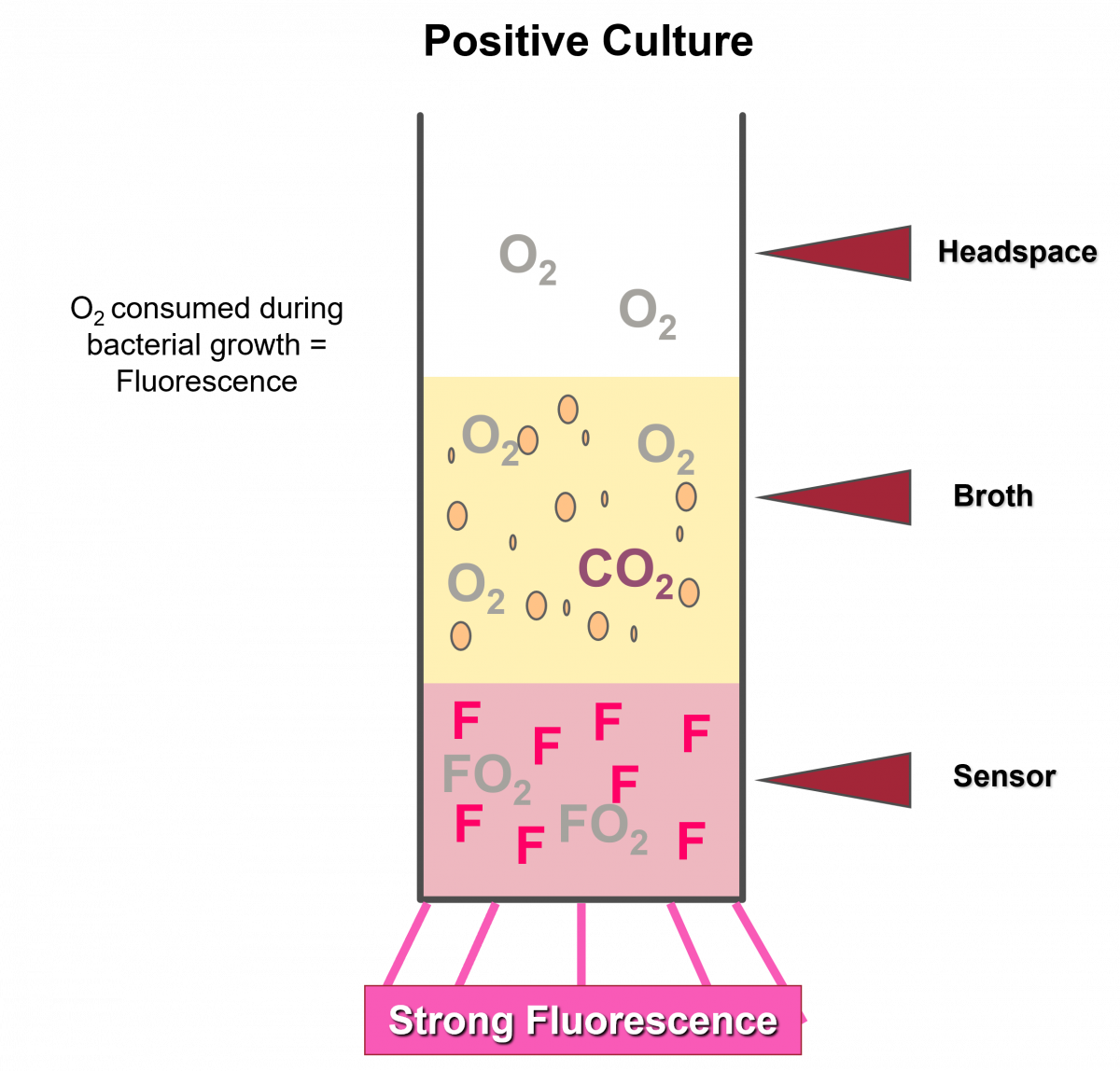
Figure 1: Positive Growth indicated by Strong Fluorescence
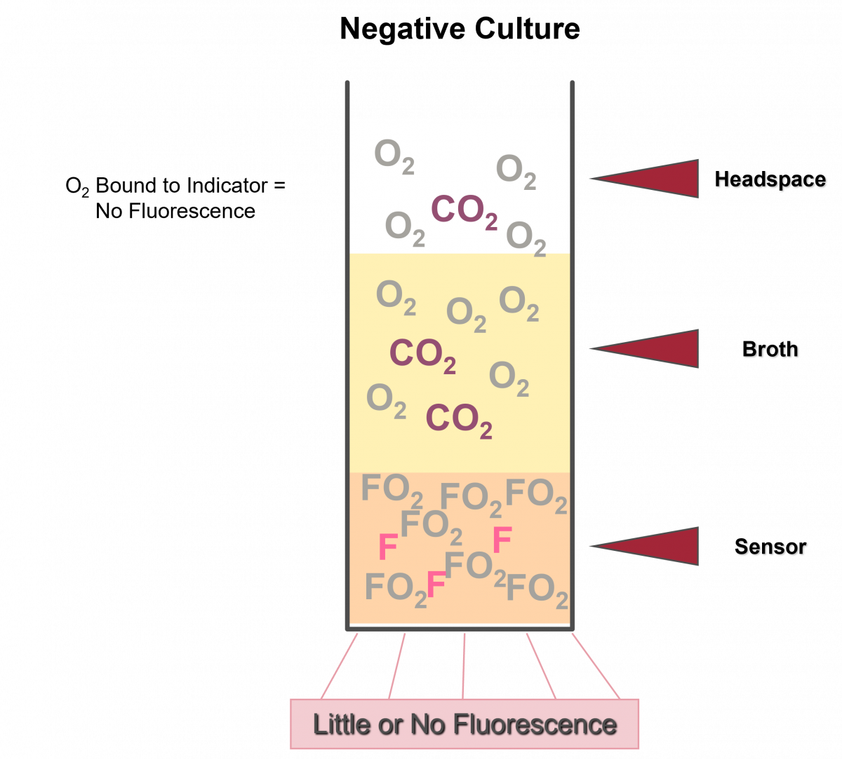
Figure 2: Negative Growth indicated by Lack of Fluorescence
Resources
Kindly provide your valuable feedback on the page to the link provided HERE
LMS Page Link
Content Creator
Reviewer
- Log in to post comments