Content Status
Type
Linked Node
Fluorescent Microscopy: Properties of a well stained slide
Learning ObjectivesDiscuss the Properties of a well stained slide in Fluorescent Microscopy. Describe staining of background and MTB (describe the background color, morphology of bacilli, colour of stain and counterstain, uniformity etc. in FM)
Light-emitting Diode Fluorescence Microscopy (LED-FM) utilises the fluorescent dye Auramine-O to stain and detect Mycobacterium species in clinical samples.
Characteristics of a Well-stained Slide
- Auramine-O stained Mycobacterium species are visible as bright yellow/ green long slender rods, slightly curved, with variable lengths, single or in clumps, with uniform staining or granular appearance against a dark background (Figure A).
-
Slides stained with Auramine-O should not have too much background fluorescence (Figure B)
Possible reasons:
- Smear thickness is not appropriate
- Insufficient decolourisation
- Counterstain too weak
- Auramine-O dries on the smear
-
Auramine-O not filtered
Possible solutions:
- Internal quality control of prepared smears and stains
- Use a stopwatch to time-staining steps
- Filter Auramine-O before use
- Add a sufficient quantity of stains to cover the smear
-
Slides stained with Auramine-O should not have pale fluorescence (Figure C)
Possible reasons:
- Smear thickness is not appropriate
- Low Auramine-O concentration
- Excessive decolourisation time
-
Stained smears exposed to daylight
Possible solutions:
- Internal quality control of prepared smears and stains
- Use a stopwatch to time-staining steps
- Store Auramine-O and stained slides in the dark
- Read stained slides as early as possible
- Non-fluorescent yellow/ green coloured bacillary shapes should not be considered as Mycobacterium species.
-
Slides stained with Auramine-O may contain stained artefacts/ background debris which are not Mycobacterium species.
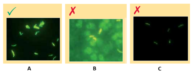
Figure: Slides stained with Auramine-O showing bright yellow/ green slender rods (A); slide with too much background fluorescence (B); slide with pale fluorescence (C); Source: Laboratory Diagnosis by Sputum Smear Microscopy
Resources
- Laboratory Diagnosis of Tuberculosis by Sputum Smear Microscopy - The Handbook, GLI, 2013.
- Manual for Sputum Smear Fluorescence Microscopy, RNTCP, CTD.
Assessment
| Question | Answer 1 | Answer 2 | Answer 3 | Answer 4 | Correct answer | Correct explanation | Page id | Part of Pre-test | Part of Post-test |
| Which of these is/are the characteristic/s of a well-stained FM slide? | Mycobacterium species are visible as bright yellow/ green long slender rods against a dark background. |
Mycobacterium species are visible as pink, long slender rods.
|
Mycobacterium species are visible as pale yellow/ green long slender rods against a dark background. | Mycobacterium species are visible as non-fluorescent yellow/ green coloured bacillary shapes. | 1 | Mycobacterium species are visible as bright yellow/ green long slender rods against a dark background. | | Yes | Yes |
Page Tags
Content Creator
Reviewer
Target Audience
- Log in to post comments
Comments
Picture of each slide can be…
Dr. Priyadarshini Wed, 10/01/2024 - 02:03
Picture of each slide can be next to the content on it.